
Diagnosis
Traumatic brain injuries can be mild, moderate or severe. If brain injury is suspected It is normally investigated even if there are no obvious signs at the time of admission.
Signs and symptoms
Signs and symptoms of mild brain injury
headache
nausea
lack of orientation
dizziness
difficulty balancing
blurred vision
fatigue
peculiar taste in the mouth
These may get worse with time and be accompanied by convulsions, slurred speech, dilation of pupils as well as weakness and numbness of limbs. This deterioration is suggestive of a more severe injury.
Signs and symptoms of moderate to severe brain injury
severe disorientation
paralysis
low heart rate
unconsciousness
respiratory depression (not breathing regularly or at all)
Signs of an open or penetrating head injury
Visible scalp or skull wound
Clear fluid running from nose (this is CSF and indicates that there is a skull fracture that may not be immdeiately obvious)
Blood or clear fulid running from ears
'Panda eyes' - this is bruising around the eyes when there is no direct injury to the eyes
Bruising behind ears
New deafness in both ears
Clinical methods of diagnosis
Neurological examination
Catagory |
Response |
Score |
Eye opening |
Spontaneous | 4 |
| To speech | 3 | |
| To pain | 2 | |
| None | 1 | |
Best verbal response |
Oriented | 5 |
| Confused | 4 | |
| Inappropriate | 3 | |
| Incomprehensible | 2 | |
| None | 1 | |
Best motor response |
Obeying commands | 5 |
| Localising | 4 | |
| Flexing | 3 | |
| Extending | 2 | |
| None | 1 |
Mild: 13 or above, LOC under 20 minutes
Moderate: GCS of 9-12, LOC of 20 minutes to 6 hours
Severe: GCS of 8 or below, LOC of 6 hours or more
If drugs or alcohol are suspected the GCS score at the time of admission may provide false results. In this case, the patient is monitored as usual and the GCS scored when the effects of substances would have worn off.
Neuroimaging
Neuroimaging is used to see if there is any obvious damage to the skull or the brain. This can include x-rays to check for fractures in minor head injuries and CT (computed tomography) scans for moderate to severe cases. Although diffuse damage can be very hard to see on scans, contusions and haemorrhage can be identified. This is very important as these factors can contribute greatly to what is known as 'secondary' injury and therefore require rapid attention, normally surgery, to minimise risks.
Monitoring the neurological responses of the patient will continue regularly to check for signs of deterioration. If the patient's condition worsens further scans maybe required and may include an MRI (magnetic resonance imaging) which can provide a clearer, more detailed picture of what is going on inside the brain. Other proceedures that may be utilised are venous dopplers and angiographies which are used to image vessels within the brain as well as EEGs which measure brain activity. In moderate to severe injuries the ICP (intracranial pressure) will also be monitored as a rise in pressure can indicate the presence of secondary injury and can, if not treated, be fatal.
Brain injuries which are classified as minor may only require a small amount of monitoring before they can go home. However, more severe injuries can require the patient to be monitored on an intensive care unit for days, weeks or months depending on the severity of the primary injury and complications related to secondary injuries. This is discussed further in the treatment and management sections of the website.
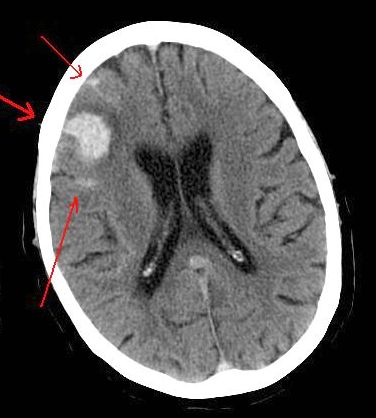
Image courtesy of Wikimedia Commons under the GNU Free Documentation License.